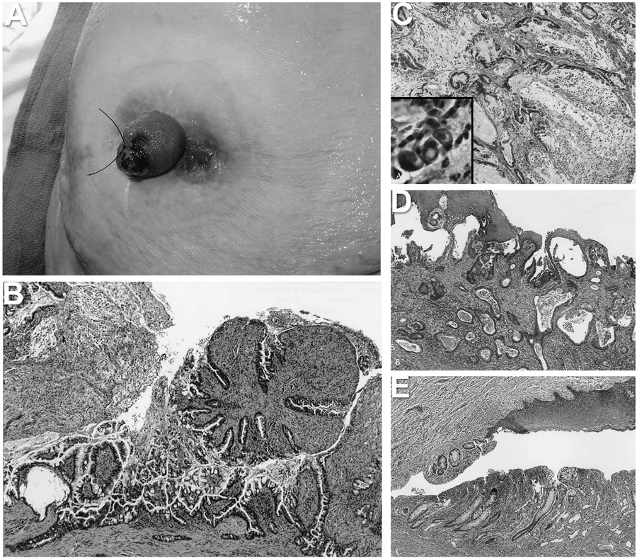
Figure 2.
Ileostomy cancer samples. A) ileostomy in situ image showing ulceration and adjacent ulcerated mass with granulation tissue; B) initial biopsy from the granulation tissue revealing proliferating epithelium with moderate cytologic atypia, thought to represent dysplasia; C) low magnification image of the tumor showing moderately differentiated carcinoma with abundant extracellular mucin pools typical of mucinous adenocarcinoma. High magnification inset showing scattered signet-ring cells infiltrating the stoma; D) section from the enterocutaneous junction showing adenocarcinoma extending underneath the squamous epithelium of the epidermis; and E) section from the enterocutaneous junction showing colonic metaplasia of the small bowel mucosa.

