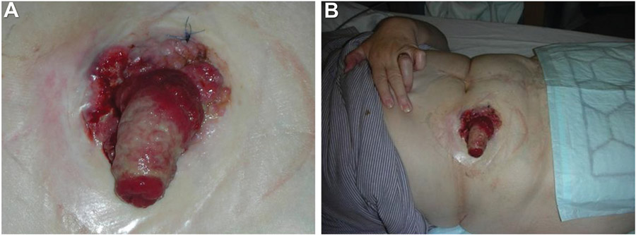
Figure 4.
A-61-year-old woman presented at the colorectal clinic with a 3-month history of decreased stoma output, weight loss, and general malaise. She also had a history of panproctocolectomy and ileostomy for ulcerative colitis at the age of 13. She had been admitted with small bowel obstruction 4 months prior to the current visit and treated conservatively. A follow-up small bowel barium follow-through did not show any small bowel obstruction. Further examination of the abdomen revealed an ulceroproliferative growth involving the mucocutaneous junction and ileostomy site extending from the 9 to 6 o’clock position (A and B). The sprout of the ileostomy site was thickened and stenosed. Biopsy from the lesion revealed an adenocarcinoma. Blood test showed a carcinoembryonic antigen level of 9, Ca 19–9 of 228, and Ca 125 of 21.6. A computerized tomography scan of her abdomen and pelvis did not show any evidence of distant metastasis. The patient underwent a wide local excision at the ileostomy site and the adjacent anterior abdominal wall with a 2-cm margin and resiting of the stoma.51 (Used with permission from the author.)

