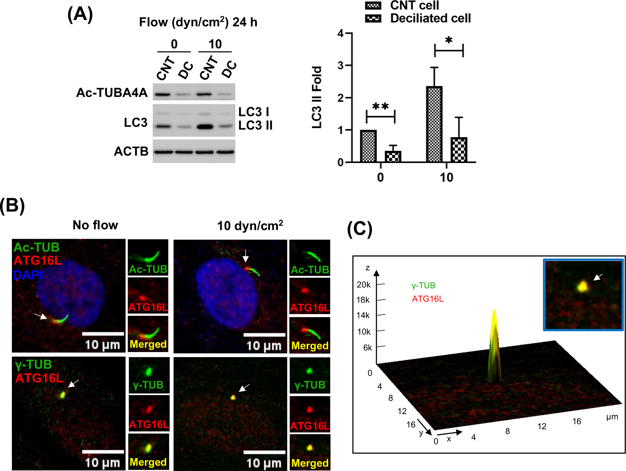
Figure 3.

PC regulate shear stress-induced autophagy in SC cells. (A) PC were disrupted by treating SC cells with 2 mM of CH for 3 days. CH was removed by switching to fresh media and SC cells were then subjected to shear stress (10 dyn/cm2) for 24 h. Protein levels of LC3-II, and acetylated TUBA4A (PC marker) were evaluated by WB. Band intensities were quantified by Image Lab™ touch software and normalized with ACTB. Data are shown as the mean ± S.D. (n=4). *, p<0.05; **, p<0.01 (Student’s t-test). CNT: control, DC: deciliated. (B) Representative immunostaining of ATG16L (red) with acetylated TUBA4A (green, upper panels) and ɣ-tubulin (green, lower panels), a maker for the basal body of PC, in SC cells in the absence or presence of shear stress for 24 h. (C) Interactive 3D surface plot analysis visualizing the colocalization of ATG16L with ɣ-tubulin (white arrows).
