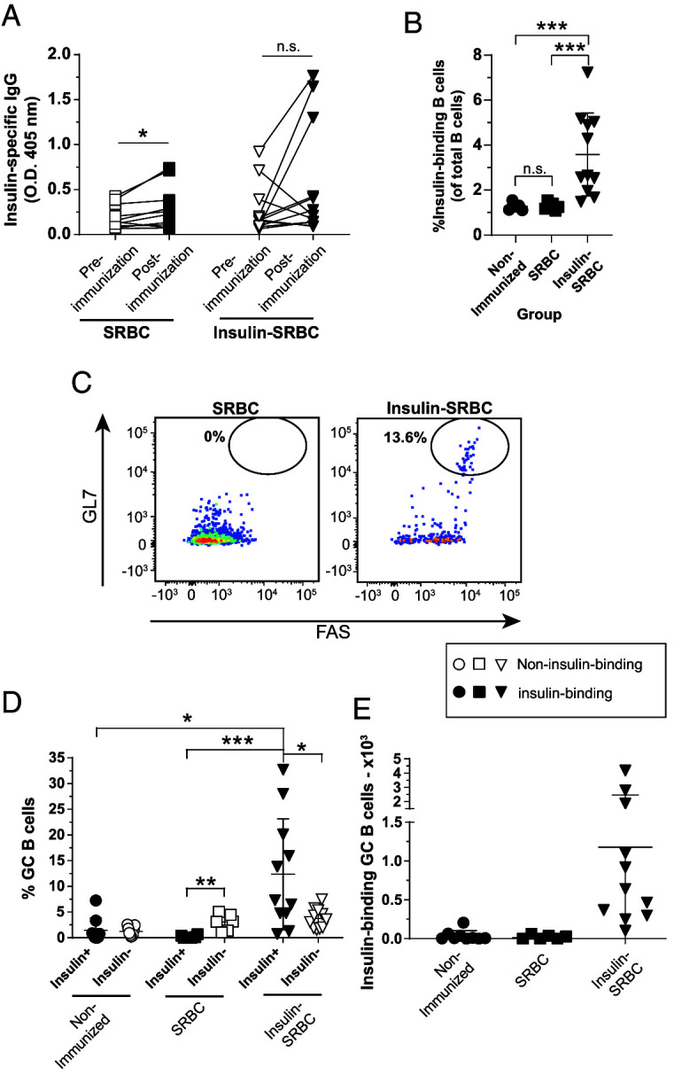
FIGURE 2.

TD immunization with insulin SRBCs elicits poor anti-insulin Ab production but drives anti-insulin GC B cell expansion.
VH125SD.NOD mice were immunized s.c. at the base of the tail with either SRBCs or insulin conjugated to SRBCs (insulin SRBCs) as described in Materials and Methods. Draining medial iliac lymph nodes and sera were harvested 10 d after immunization. (A) Serum anti-insulin Ab production was measured by ELISA in VH125SD.NOD mice before and after immunization with either insulin SRBC or SRBC. The frequency of (B) insulin-binding B cells among total B cells after immunization with either SRBC or insulin SRBCs. (C) Representative flow cytometry plots identify GC B cells as in Fig. 1 among insulin-binding B cells in draining lymph nodes of mice immunized with SRBCs (left) and insulin SRBCs (right). The (D) frequency (E) and number of GC B cells among insulin-binding B cells between nonimmunized, SRBC-, and insulin SRBC–immunized mice are shown for mice that had >20 insulin-binding B cells in the parent gate. Eight- to fourteen-wk-old male and female NOD mice were used. n ≥ 6 mice per group, n ≥ 2 experiments. *p < 0.05, **p < 0.01, ***p < 0.001, paired two-tailed t test (A) and Mann-Whitney U test (B).
