. 2023 Aug 22;15(1):2245111. doi: 10.1080/19420862.2023.2245111

Figure 2.

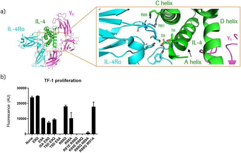
In panel A, the crystal structure of IL-4 bound to the IL-4 receptor complex is shown. A zoomed-in representation on the right side shows the location of IL-4 helices A, C, & D, and the positions of relevant amino acids that interact with the receptor. In panel B, a bar graph shows the relative activities of different IL-4 mutants. The mutations E9Q, R88Q, and R88Q W91A result in very similar activities compared to non-mutated IL-4. E9A, I5A E9Q, T6D E9Q, and R88A show reduced activities, and R88Q N89A shows very little activity. T6D E9A, R81E R88Q, and K84E R88Q show no detectable activity.

Inactivation of targeted split IL-4 generates prodrug modules.

(a) Structural depiction of the IL-4/IL-4Rα/common gamma chain (γC) ternary complex, PDB ID 3BPN.34 Key residues in IL-4 that interact with IL-4Rα are indicated in green. (b) Effect of IL-4 mutations (generated in the context of CD38-targeting 3+1 split IL-4 PACE educts) on IL-4 signaling activity, as measured by proliferation of TF-1 cells treated with 100 nM of the respective molecules. TF-1 cells express CD38 – see following section for more details.