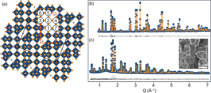
Figure 1.
(a) Schematic of the NaNb13O33 crystal structure highlighting the (5×3–2)∞ blocks of NbO6 octahedra and each unique Nb site. (b) Neutron powder diffraction of 1-NNO obtained at 300 K, λc = 1.5 Å. (c) Synchrotron X-ray powder diffraction of 1-NNO at 300 K, λc = 0.45788 Å. The diffuse background feature is due to the Kapton capillary sample holder. The inset shows an SEM image of pristine 1-NNO particles

