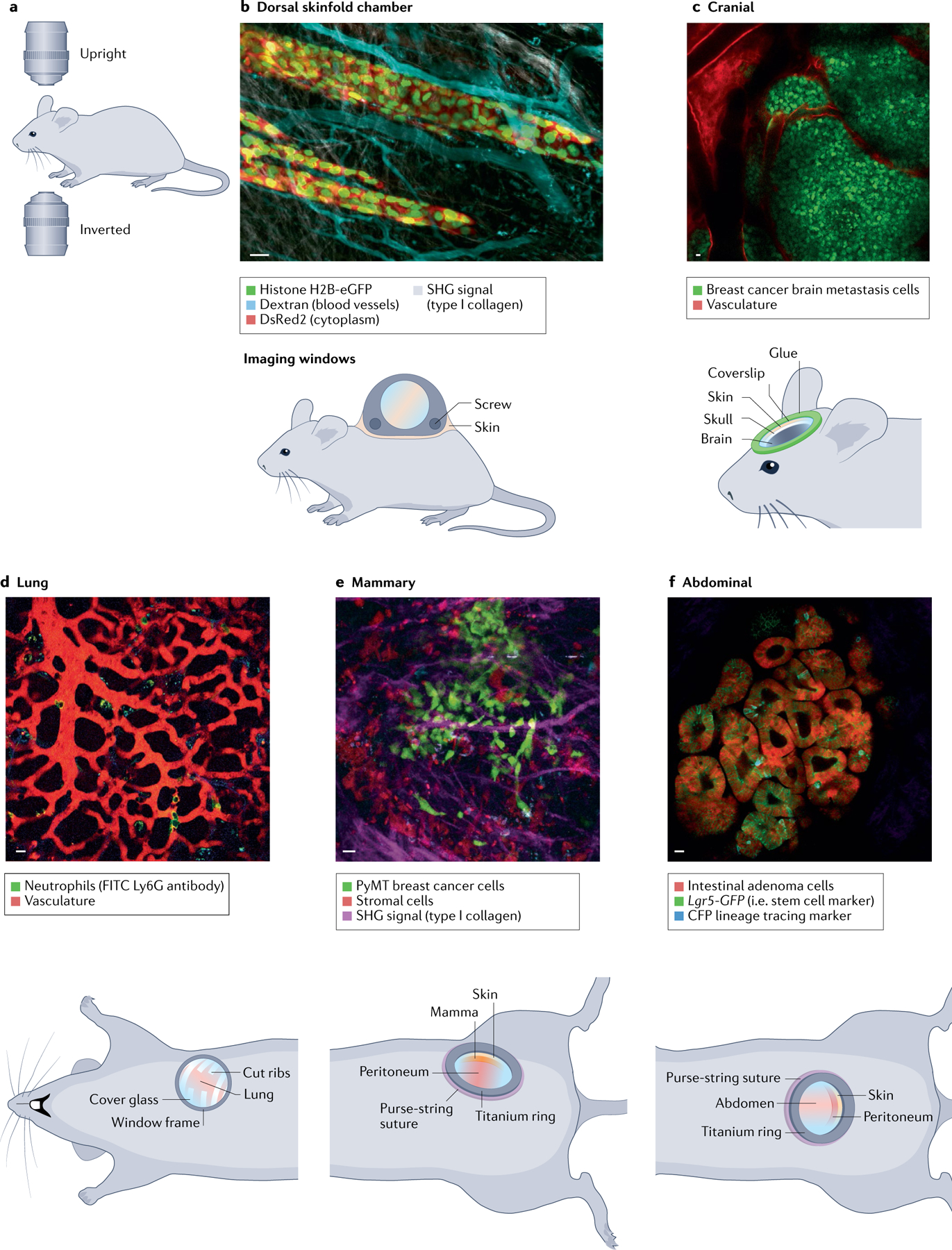
Fig. 2 |. Intravital microscopy of tissues through optical imaging windows.

Murine models can be imaged with upright or inverted microscopes (part a). Examples of chamber types (cartoons) and representative example multicolour intravital microscopy (IVM) images taken through the imaging windows of the dorsal skinfold chamber (part b), cranial window (part c), lung window (part d), mammary imaging window (part e) and abdominal imaging window (part f). Scale bars represent 20 μm. eGFP, enhanced green fluorescent protein; SHG, second harmonic generation. Part b (IVM image) reprinted with permission from REF.384, Rockefeller University Press.
