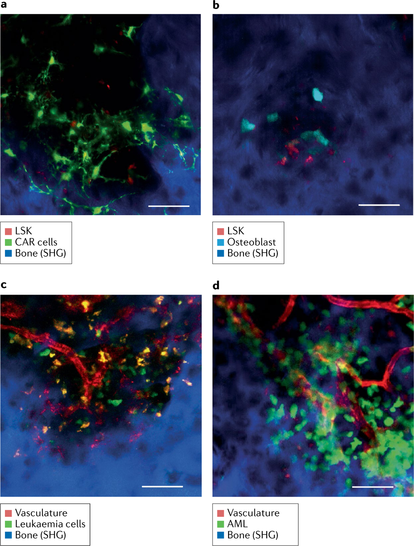
Fig. 5 |. Bone marrow imaging in situ in the leukaemia mouse model.

Representative intravital two-photon skull images. Bone tissue was detected by second harmonic generation (SHG), which captures collagen molecules without staining (represented in blue). Scale bar: 50 μm. a,b | Images of mouse LIN−SCA1+KIT− (LSK) cells within the bone marrow niche: mouse LSK-tdTomato cells transplanted into Cxcl12-GFP recipient mouse (green, CXCL12-abundant reticular (CAR) cells; red, LSK cells) (part a); and mouse LSK-tdTomato cells transplanted into Col1a1 2.3-CFP mouse (cyan, osteoblasts; red, LSK cells) (part b). c | Intravital image of a murine chronic myeloid leukaemia model made by transplantation of leukaemia cells (BCR-ABL-ires-GFP transfected LIN−SCA1+KIT+ cells). Green, leukaemia cells; red, blood vessels. d | Intravital image of a murine acute myeloid leukaemia (AML) model made by transplantation of leukaemia cell line (C1498-eGFP). Green, leukaemia cells; red, blood vessels.
