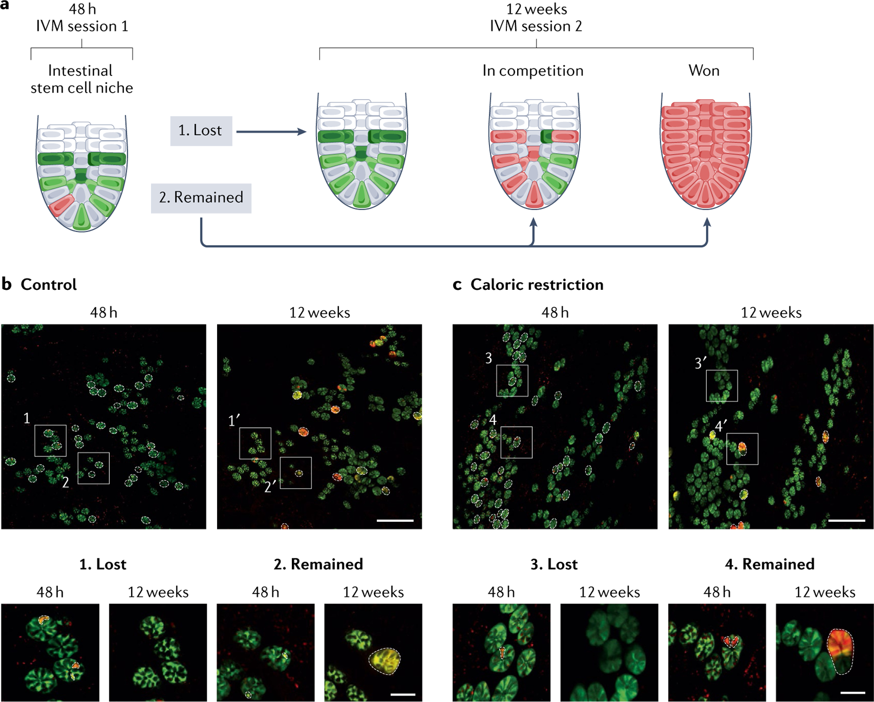
Fig. 6 |. Intravital microscopy of calorie restriction reveals slower and stronger stem cell competition.

a | By repeated intravital microscopy (IVM) of the same crypts 48 h and 12 weeks after fluorescent labelling of the stem cells, stem cell competition can be visualized. Stem cell clones are either lost or remain within the crypts, represented by the presence of red cells. b,c | IVM images of the same intestinal areas in control conditions (part b) and upon caloric restriction (part c). Dotted lines highlight the crypts in which labelled stem cell clones were present at 48 h and 12 weeks after stochastic fluorescent labelling. Zoomed-in images (bottom panels) show examples of clones that were lost (1 and 3) or remained (2 and 4) in both conditions. Scale bars represent 250 μm (top panels) or 50 μm (bottom panels). Adapted with permission from REF.279, Elsevier.
