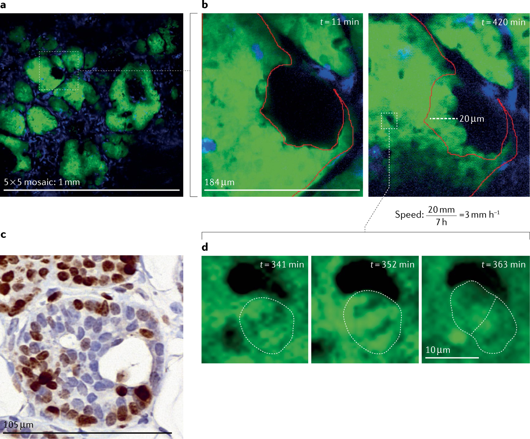
Fig. 7 |. Intravital microscopy of the ductal carcinoma in situ in the PyMT mouse model.

a | Large-volume high-resolution intravital microscopy (IVM) of the ducts provides an overview of the tissue and allows identification of the tissue stage and of incompletely filled ducts (white box). A 5 × 5 mosaic of the mammary gland imaged through a mammary imaging window implanted in a transgenic mouse model that expresses the PyMT oncogene and the fluorescent proteins Dendra2 in the mammary epithelium (green) and CFP in macrophages (blue). b | Stills from a continuous time-lapse movie of the duct showing migration of the cells over 7 h imaging duration. Red outline indicates position of cells at time t = 0 min. After only a brief imaging period (left), displacement of the cells is not obvious. However, after 7 h of imaging (right), cells have moved appreciably. Dashed line indicates the distance the cells have moved and can be used to calculate the speed of cell movement during imaging. c | Ki67 staining (brown) of fixed tissues taken from another mouse shows ~60% of cells are actively proliferating in similar structures. d | High-resolution stills from the IVM movie captures the division of a single cell (white dashed outline drawn by hand indicates the boundary of the dividing cell and the daughter cells), as evidenced by chromosomal separation over ~30 min.
