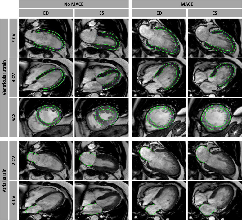
Figure 1.
Cardiovascular magnetic resonance-feature tracking (CMR-FT). CMR-FT analyses in a patient with and without a major adverse cardiac event (MACE). Ventricular analyses were performed in long-axis 2- and 4-chamber views (CV) as well as in short-axis (SAX) stacks including basal, mid-ventricular, and apical slices, with only an exemplary mid-ventricular slice illustrated in this figure. Atrial strain analyses were performed in long-axis 2- and 4 CV images as well. Myocardial border delineations are presented in end diastole (ED) and end systole (ES).

