. 2023 Aug 11;32(3):e007523. doi: 10.1590/S1984-29612023047

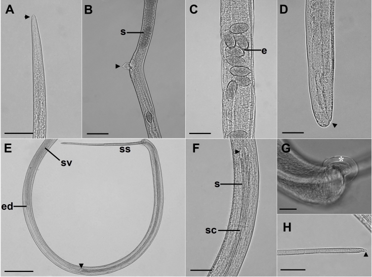
Figure 2. Microscopic views of Capillaria cairina n. sp. (A) Female anterior end (arrowhead). Scale bar = 50 µm; (B) Pre-equatorial region, lateral view, where the stichocytes (s) end and the vulva with its vulvar appendix (arrowhead). Scale bar = 100 µm; (C) Uterus with eggs (e) with clearly visible polar plugs. Scale bar = 50 µm; (D) Posterior extremity, lateral view; the anal opening is observed (arrowhead). Scale bar = 50 µm; (E) Posterior extremity of the male, lateral view; the base of the spicule (arrowhead) and extruded spicular sheath (ss) can be observed. Scale bar = 200 µm; (F) Base of the spicule (arrowhead). Scale bar = 50 µm; (G) Posterior extremity of the male, lateral view, lobe with a delicate membrane (*). Scale bar = 20 µm; (H) Distal end of the spicule with a rounded tip and the presence of a thin membrane at its apex (arrowhead). Scale bar = 50 µm. s = spicule; ss = spicular sheath; sc = spicular canal; ed = ejaculatory duct; sv = seminal vesicle.

Figure 2