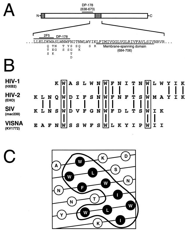
FIG. 1.
Sequence conservation and predicted structure of the tryptophan-rich region. (A) Schematic representation of gp41, showing the location and sequence variability of the tryptophan-rich region and the overlapping sequences of the 2F5 epitope and DP-178 peptide. Shaded regions indicate the N-terminal fusion peptide and the membrane-spanning domain. The amino acids in the HIV-1 sequence database which occur at each position are denoted underneath the respective amino acid in the HXB2 strain. (B) Alignment of the tryptophan-rich sequence of HIV-1 HXB2 gp41 with homologous regions of HIV-2, SIV, and visna virus glycoproteins. Conserved tryptophan residues are boxed. (C) Helical net projection of the tryptophan-rich region. The outlined region indicates the cluster of conserved hydrophobic residues shown in black.

