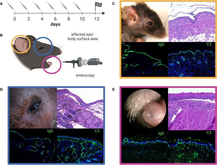
Figure 5.
Anti-laminin α3 mucous membrane pemphigoid (MMP) mouse model. Rabbit anti-murine laminin α3 IgG is injected subcutaneously (s.c.) into adult C57Bl/6 mice every other day over a time period of 10 days (A). The clinical manifestation of the mouse model seen on experimental day 12 can be quantified by the use of a validated scoring system comprising the affected body surface area (yellow, C), the affected eye-area (blue, D), and the severity of oral lesions as examined by endoscopy (pink, E) (B). The color-framed boxes (C-E) show the clinical presentation (upper left panel), H&E stained lesional histopathology with an inflammatory infiltrate and split formation of the dermal-epidermal/epithelial junction (upper right panel) and, linear deposits of IgG (lower left panel) and C3 (lower right panel) along the basal membrane zone by direct immunofluorescence microscopy. Lesions, crusts and erosions of the skin are mostly restricted to the head, neck, and upper back of mice (C). The image was created with BioRender.com.

