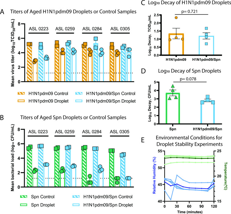
Fig 2.
Stability of S. pneumoniae and influenza viruses in droplets. (A–D) Viral and bacterial loads of H1N1pdm09 and Spn were assessed after exposure of 10 × 1 µL droplets to 43% relative humidity (RH) at room temperature for 2 h. Microbes were suspended in ASL from four different HBE cell donors as indicated in A and B. Control loads were determined using 10 µL of bulk solutions in closed tubes at room temperature. (A) The stability of H1N1pdm09 in droplets containing H1N1pdm09 or H1N1pdm09/Spn measured by TCID50 assay, and (C) log10 decay for each individual ASL culture were determined. (B) The stability of Spn in droplets containing Spn or H1N1pdm09/Spn measured by CFU assay, and (D) log10 decay for each individual ASL culture were determined. Differences were assessed using Welch’s unpaired t-test. (E) The RH and temperature were recorded every 15 min during stability experiments. Temperature (light green) and RH (light blue) for each ASL replicate are shown. The average temperature (dark green) and RH (dark blue) for all experiments are also included. Bacterial samples with no detection were placed at 1/2 the LOD.

