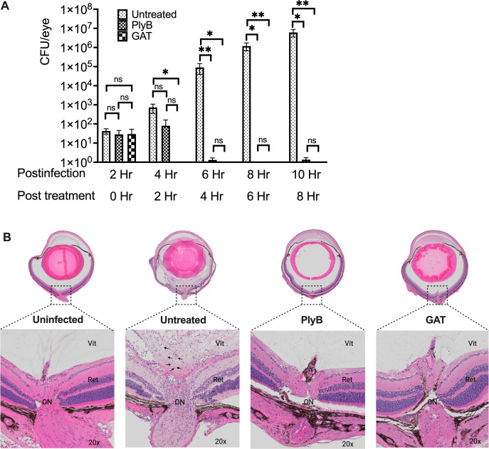
Fig 7.
PlyB sterilized eyes in experimental B. cereus endophthalmitis. Treatment with PlyB or GAT prevented bacterial growth and preserved ocular structure. C57BL/6J mice eyes were infected with 100 CFU B. cereus. After 2 hours post-infection, eyes were treated with 420 µg/mL PlyB or 250 µg/mL GAT. Untreated, infected, and treated globes were harvested and analyzed at 0, 2, 4, 6, and 8 hours post-treatment. (A) PlyB and GAT completely reduced the B. cereus load by 4 hours post-treatment. Compared with untreated eyes, bacterial load was significantly low (*P ≤ 0.05, **P ≤ 0.01) in PlyB and GAT-treated eyes at 4, 6, and 8 hours post-treatment. Values represent means ± SEM of n ≥5 eyes at each time point with at least two independent experiments. *P ≤ 0.05, **P ≤ 0.05, and ns P ≥ 0.05. (B) Harvested eyes were processed for hematoxylin and eosin staining. At 8 hours post-treatment, bacteria were found in untreated eyes (black arrow bottom panel). Untreated eyes were severely inflamed with inflammatory cells near the optic nerve area. The retina was partially detached, and fibrin deposition was observed. In contrast, intact retinal layers and minimal inflammation with no sign of bacterial presence were observed in PlyB- and GAT-treated eyes. Sections represent two eyes per time point with at least two independent experiments: original magnification top panel ×10, bottom panel ×20. B. cereus is denoted by black arrows. Ret, retina; Vit, vitreous; AqH, aqueous humor; Ir, iris; L, lens.

