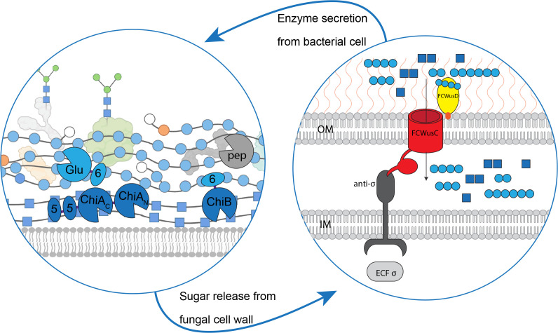
Fig 5.
Proposed pathway of synergistic action by the proteins of the FCWUL. Left panel: model of a fungal cell wall undergoing hydrolysis by FCWUL enzymes. Right panel: theoretical model of the FCWUL (OM, outer membrane of bacterial cell; IM, inner membrane of bacterial cell). Components are not shown to scale in either panel. All FCWUL enzymes are depicted as being extracellular, because they all carry standard bacterial SpI signal peptides (19 - 21) and (except for CpGlu16A) the C-terminal domain for secretion via the type IX secretion system, T9SS (22). Dark blue squares, GlcNAc; light blue circles, β-1,3-linked Glc; orange circles, β-1,6-linked Glc; white circles, terminal Glc; green circles, mannose found on glycosylated FCW proteins.

