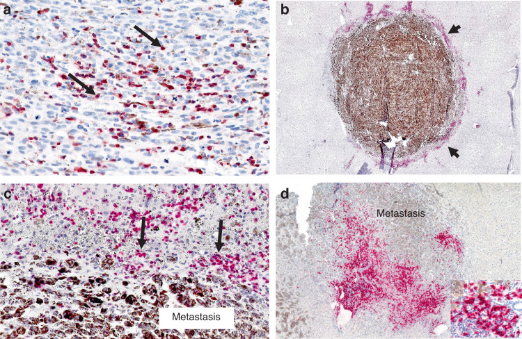
Fig. 3. Immunohistochemistry showing expression of lymphocyte markers in primary uveal melanoma and uveal melanoma liver metastases.
a Primary uveal melanoma. Moderate CD3 TILs (arrows) expression (red chromogen) (score = 2; 5–50%). b Uveal melanoma liver metastasis. Intense peri-tumoral CD3 infiltrate (arrowheads) (red chromogen) (score = 3; >50%) surrounds heavily pigmented metastasis. c Higher magnification of (b). Arrows highlight CD3+ peri-tumoral lymphocytes. d Uveal melanoma liver metastasis. Prominent peri-tumoral CD20 infiltrates (red chromogen). Inset (right lower) shows CD20+ lymphocytes.

