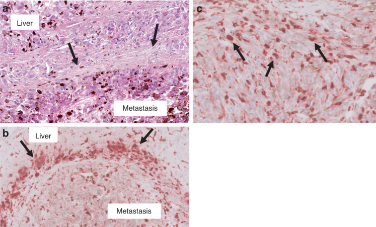
Fig. 4. Immunohistochemistry showing expression of macrophage markers in uveal melanoma liver metastases.
a Uveal melanoma liver metastasis with desmoplastic histopathological growth pattern (HES stain). The metastasis (lower half of this image) is separated from the liver (upper part of the field) by a distinct thickened band of desmoplastic collagen (arrows). b Peri-tumoral band-like CD163 infiltrate (arrows) (reddish-brown chromogen) associated with the desmoplastic annulus, as seen in (a). c Uveal melanoma liver metastasis. CD68+ tumour-infiltrating macrophages (arrows) present within the metastasis (score =3; >50% of tumour area contains these TIMs).

