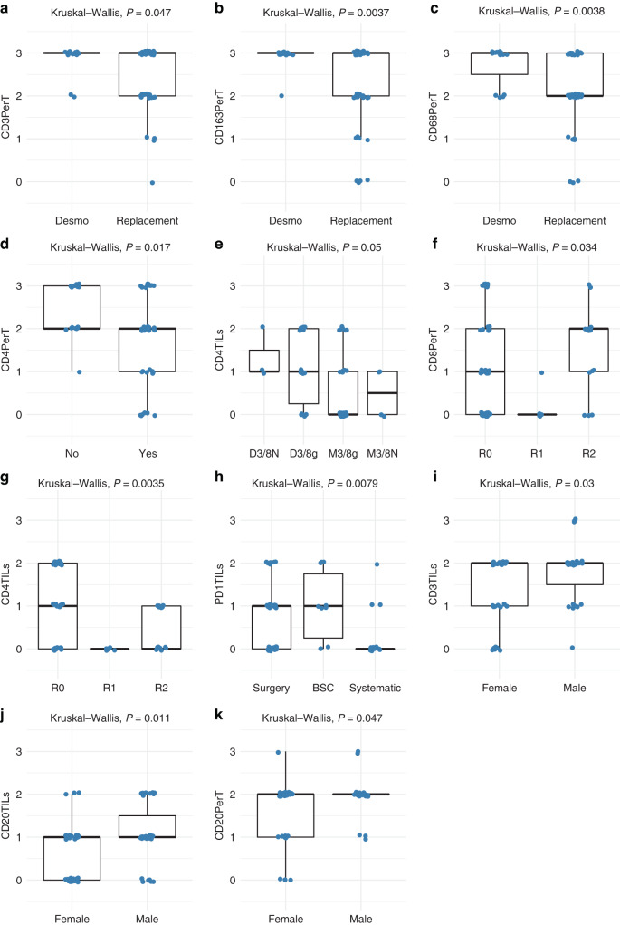
Fig. 5. Boxplots showing significant differences for immune cell infiltrates in uveal melanoma liver metastases analysed for various prognostic factors.
a–c Uveal melanoma liver metastases stratified according to predominant desmoplastic or replacement histopathological growth pattern, d BAP1 loss, e aCGH risk group, f, g resection margins, h treatment, and i–k and gender. The immune parameters for which significant differences were noted are indicated along the Y-axis (Kruskal–Wallis test).

