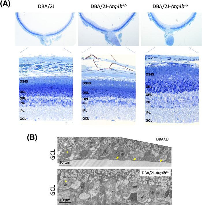
Fig. 2. Atg4b deletion alters retinal morphology in DBA/2J mice.
A Cross-sectional histological images showing changes in retinal morphology in DBA/2J-Atg4bko mice, including increased thickness of the INL and OS, and disorganization of the OS and IS (higher magnification shown in SM, Fig. 2). Images are representative of at least 6 animals per group. OS outer segment, IS inner segment, ONL outer nuclear layer, OPL outer plexiform layer, INL inner nuclear layer, IPL inner plexiform layers, GCL ganglion cell layer. B Ultrastructural images of the GCL showing the higher RGC density (asterisk, representative) and the absence of apoptotic and necrotic bodies (arrows) with deletion of Atg4b.

