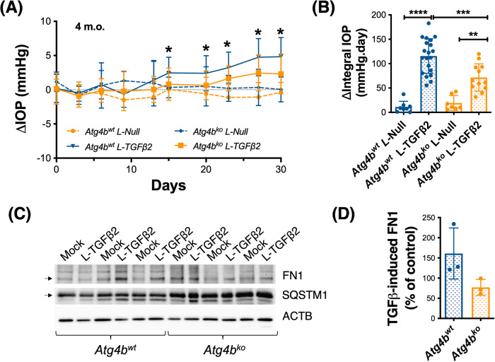
Fig. 6. Atg4bko deficiency protects against TGFβ2-induced IOP elevation and fibrosis.
Atg4bko and Atg4bwt mice (4 m.o.) were intravitreally injected in the right eye with L-TGFβ2 or L-Null virus (2 × 10⁶ TU/eye, 2 μL bolus). The contralateral eye remained non-infected. Nocturnal IOP was monitored twice per week. A Mean ΔIOP (mm Hg) calculated between the injected and the mock fellow eye monitored over time. IOP exposure was calculated and plotted on (B). *p < 0.05, **p < 0.01, ***p < 0.001, ANOVA with Bonferroni post hoc test (Atg4bwt L-Null n = 8, Atg4bwt L-TGFβ2 n = 21; Atg4bko L-Null n = 8, Atg4bko L-TGFβ2 n = 12). C WB quantification of fibronectin (FN1) and SQSTM1 protein expression levels in dissected iridocorneal region. ACTB was used for normalization. D Quantification of TGFβ2-induced FN1 protein levels from the densitometric analysis of the bands.

