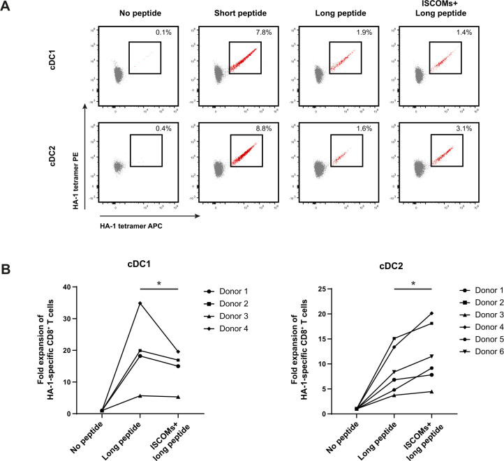
Figure 3.
Enhanced antigen cross-presentation in cDC2s upon ISCOMs. (A) HA-1- specific CD8+ T-cell expansion indicated by double-positive HA-1 tetramer staining (in red) for cDC1s and cDC2s after stimulation with short peptide, long peptide, or ISCOMS+ long peptide. DCs (100,000 cells) were incubated with PBMCs (750,000 cells) in a 1:7.5 ratio. Representative flow cytometry data are shown from one healthy DC donor. (B) Fold expansion of HA-1-specific CD8+ T cells in cDC2s and cDC1s. The numbers of different healthy DC donors used are depicted in the graphs. Statistical analyses were done using paired non-parametric t-test. *p<0.05. cDC1, conventional type 1 DC; cDC2, conventional type 2 DC; ISCOMs, immune stimulatory complexes; PBMCs, peripheral blood mononuclear cells.

