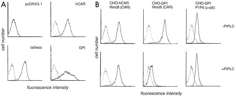
FIG. 2.
Expression of CAR on transfected CHO cells. (A) Flow cytometry. CHO cells transfected with cDNA encoding wild-type hCAR or truncated CAR (tailless and GPI), and control cells transfected with the empty pcDNA3.1 vector, were incubated first with MAb RmcB (heavy line) or with a control antibody (MOPC 195 [thin line]) and then with fluorescein isothiocyanate-conjugated goat antibody to mouse immunoglobulin. All panels are shown on the same scale. (B) Flow cytometry after PIPLC treatment. Transfected CHO cells were incubated for 30 min at 37°C in RPMI 1640 supplemented with 0.2% bovine serum albumin, 50 μM 2-mercaptoethanol, 10 mM HEPES (pH 7.0), and 0.1% sodium azide, with or without the addition of PIPLC (0.4 U per million cells; Sigma). Cells were then washed and stained with MAb RmcB, MOPC 195, or MAb P1F6, which recognizes the integrin αvβ5. All panels are shown on the same scale. CAR expression on GPI-CHO cells was reduced 30-fold after PIPLC treatment.

