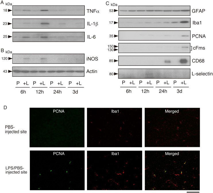
Figure 2.
Induction of inflammatory cytokines in LPS-injected rat brain
A. Induction of inflammatory cytokines. PBS was injected into the left cerebral cortex and LPS/PBS was administered in the right cerebral cortex (see Material and methods). Both areas were cut out at 6, 12, and 24 h, and at 3 days post-LPS injection, and the tissue extracts were prepared as described in Material and methods. The tissue extracts (20 μg protein) of the PBS (vehicle)-injected site (P) and the LPS/PBS-injected site ( + L) were immunoblotted for TNFα, IL-1β, and IL-6. A representative result is shown.
Molecular size (kDa) is indicated on the left side.
B. Induction of iNOS. The same samples as in A were immunoblotted for iNOS and actin. A typical result is shown.
C. Induction of glial markers, proliferation marker, monocyte/macrophage marker, and neutrophil marker. The same samples as in A were immunoblotted for GFAP, Iba1, PCNA, cFms, CD68, and L-selectin. A typical result is shown.
D. Immunohistochemical detection of proliferating cells in the LPS/PBS-injected region. Cryosections were cut from the brain into which LPS had been injected 3 days prior and stained dually with anti-PCNA antibody (PCNA; Green) and anti-Iba1 (Iba1; red) antibody. PBS-injected and LPS/PBS-injected sites are shown at the upper and lower positions, respectively. Merged images are shown on the right side. Scale bar = 50 μm.

