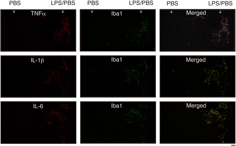
Figure 3.
Detection of inflammatory cytokines by immunohistochemical staining
coronal sections were prepared from the brain in which PBS (vehicle) and LPS/PBS had been injected 12 h prior, as shown in Figure 1B. The cryosections were dually stained with anti-TNFα antibody and anti-Iba1 antibody (top), anti-IL-1β antibody and anti-Iba1 antibody (medium), and anti-IL-6 antibody and anti-Iba1 antibody (bottom). Merged images are shown on the right side. Cytokine and Iba1 can be seen as red (Alexa Fluor-568) and Green (Alexa Fluor-488), respectively. Scale bar = 500 μm.

