Figure 4.
Ability of glial cells to induce inflammatory cytokines
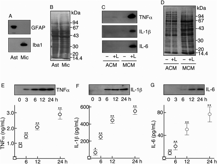
A. Purity of astrocytes and microglia. The cell extracts of astrocytes and microglia were immunoblotted for GFAP and Iba1.
B. Protein profile of glial cell extract. Cellular extracts of astrocytes and microglia were subjected to SDS-PAGE and transblotting. The transblotted PVDF membrane was stained with 0.1% Coomassie Brilliant Blue (CBB).
C. Detection of inflammatory cytokines. Two astrocytic cultures and two microglial cultures were prepared. Each culture was stimulated with LPS (0.5 μg/mL) ( + L) and the others remained as nonstimulated control (–). At 24 h, each medium of astrocytes and microglia was recovered as astrocytic conditioned medium (ACM) and microglial conditioned medium (MCM), respectively. Two ACMs (–, + L) and two MCMs (–, + L) were concentrated, freeze-dried, and immunoblotted for each of TNFα, IL-1β, and IL-6.
D. Protein profiles of ACM and MCM. Two ACMs (–, + L) and two MCMs (–, + L) were subjected to SDS-PAGE and transblotted to PVDF membrane. The membrane was stained with Coomassie Brilliant Blue (CBB).
E, F, G. Determination of inflammatory cytokines in LPS-stimulated microglia. Five microglial cultures (2 × 10⁶ cells) were prepared and then stimulated with LPS (0.5 μg/mL) as described in the Material and methods. At 0, 3, 6, 12, and 24 h, the medium was recovered and the media were concentrated, freeze-dried, and immunoblotted for TNFα (E), IL-1β (F), and IL-6 (G). At the same time, the medium taken from each culture was used to determine TNFα (E), IL-1β (F), and IL-6 (G) levels by ELISA.

