Fig. 1.
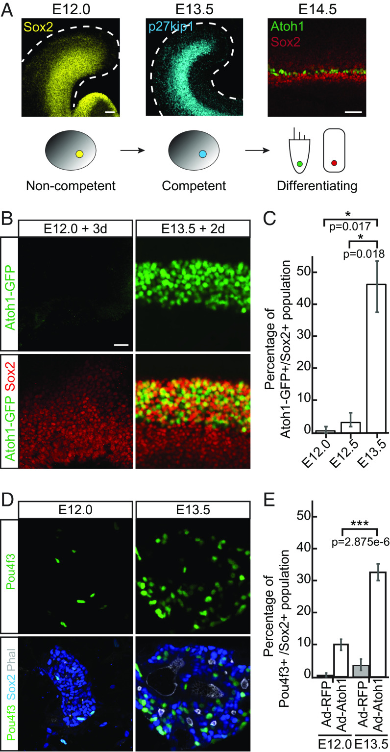
Prosensory progenitors in the cochlear duct acquire competence to differentiate as hair cells and supporting cells between E12.0 and E13.5. (A) Diagram demonstrates early embryonic development of the organ of Corti. At E12.0, Sox2-positive proliferating progenitors (yellow) start to exit cell cycle in a wave spreading from the apex toward the base of the cochlea. By E13.5, most progenitors become postmitotic and express high levels of p27kip1 (cyan). At E14.5, Atoh1-positive hair cells (HCs, green) and the surrounding Sox2-positive supporting cells (SCs, red) are specified. (Scale bar, 50 μm.) (B) Representative immunofluorescent images of the whole cochleae isolated from E12.0 and E13.5 Atoh1-GFP transgenic reporter animals and harvested for characterization after 3-d or 2-d in culture, respectively. GFP-positive hair cells (green) and Sox2-positive supporting cells (red) are labeled. (Scale bar, 20 μm.) (C) Quantitative analysis of the cultures in B demonstrates activation of Atoh1-GFP reporter in E12.0 explants (0.8%; n = 6), E12.5 explants (3.6%; n = 3) and E13.5 explants (47%; n = 3). (D) Representative immunofluorescent images show the progenitor cells isolated from the cochlear ducts at E12.0 and E13.5, infected with Ad-RFP-control or Ad-Atoh1-RFP virus, and maintained in culture for 3 d. Note that only in E13.5 cultures Atoh1 overexpression results in formation of the sensory rosettes with a small lumen formed by the actin-rich (Phalloidin, white) apical surfaces of the polarized Pou4f3–positive hair cells (green) and surrounding Sox2-positive supporting cells (blue). (Scale bar, 20 μm.) (E) Bar graph shows the quantitative analysis of cultures in D. Compared to other conditions, a significant increase in the percentage of Pou4f3-positive hair cells is observed in the E13.5 cultures where Atoh1 was overexpressed (n = 3 for each condition).

