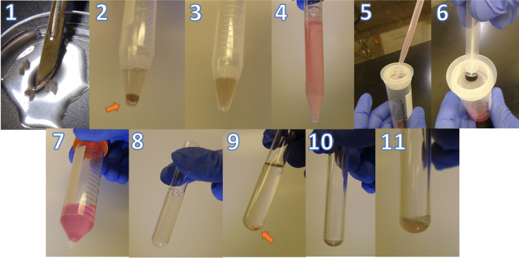
Figure 4. Representative images of retina digestion protocol.
1. Diced retinas using a scalpel. 2. Retinas resuspend in digestion buffer. 3. Digested retinas after incubation for 30 min at 37 °C. 4. Digested retinas after adding 10 mL of DMEM + 10% FBS. 5. Straining the suspension through a mesh. 6. Using the rubber end of a syringe plunger to gently press the samples through the mesh. 7. Cell pellet after centrifugation. 8. Cell suspension in a 5 mL FACS tube. 9. Cell pellet after spinning down. 10. Remaining pellet in 100 μL of PBS after aspiration. 11. Resuspended cells ready for staining.

