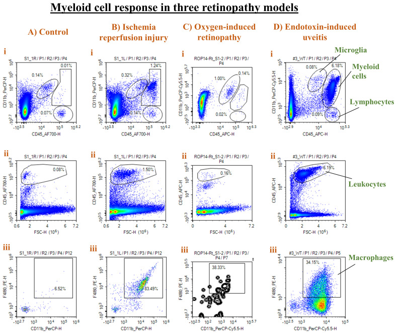
Figure 6. Representative results from three different retinal injury models.
Panels from control (A), ischemia-reperfusion (IR) injury (B), oxygen-induced retinopathy (OIR) (C), and endotoxin-induced uveitis (EIU) (D) show density plots of CD11b vs. CD45 to distinguish microglia from myeloid cells and lymphocytes (i), CD45hi leukocytes (ii), and F4/80+ CD11b+ macrophages as a percentage of CD45hi cells (iii). A strong leukocyte and macrophage infiltration was observed in both the IR and EIU models, with a stronger response in the EIU model. On the other hand, the OIR model was associated with more microglial (CD11b+ CD45low) proliferation.

