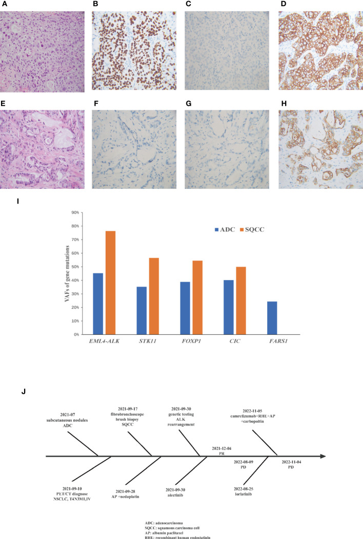
Figure 2.
A comparison of the Hematoxylin and eosin staining results from the metastatic subcutaneous nodules and primary lung tumor. Immunohistochemical show that the lung tumor biopsy is (A) squamous cell cancer (B) positive for p40 (C)negative for TTF1 (D) positive for CK7, and metastatic subcutaneous nodules are (E) adenocarcinoma (F) negative for TTF1 (G) negative for Napsin A (H) positive for CK7. (I) Variant allele frequencies (VAF) of gene mutations in different morphological sites. (J) Flow chart of patient treatment.

