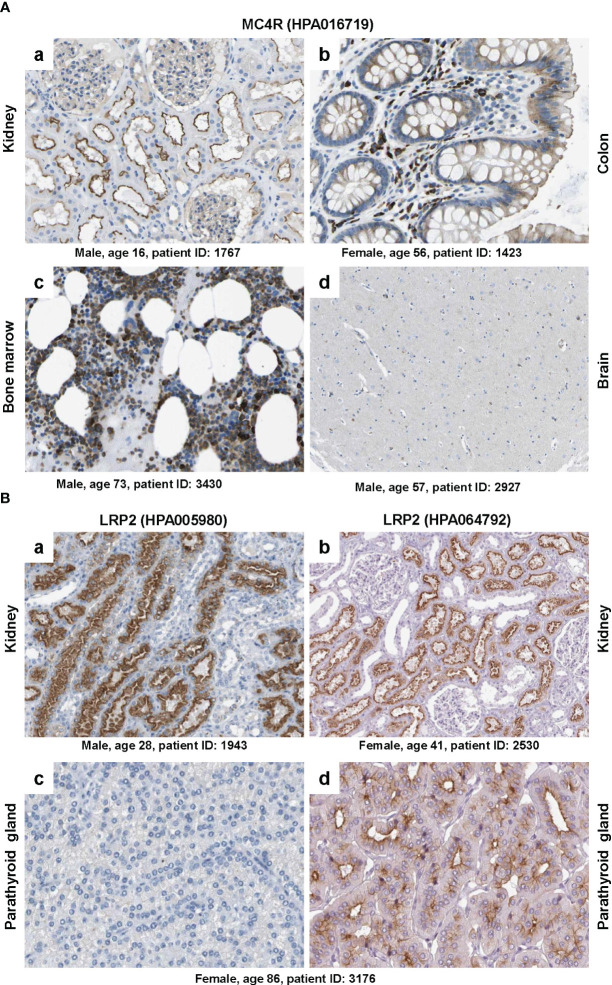
Figure 5.
Immunohistochemical staining of MC4R and LRP2/megalin. (A) Tissue sections from (a) kidney, (b) colon, (c) bone marrow and (d) brain were stained with a validated antibody (HPA016719) specific for MC4R. Please note that the antibody stained tubular cells in the kidney, hematopoietic cells in the bone marrow, glandular cells in the colon, while only showing low staining in some neuronal cells in the brain. (B) Tissue sections from (a, b) kidney and (c, d) parathyroid gland were stained with two (validated) polyclonal rabbit antibodies (i.e., HPA005980 and HPA064792) directed against human LRP2. Please note that both antibodies stained similar structures in kidney, while the staining was markedly different in two sections of the parathyroid gland that were taken from the same patient. All images were taken from the Human Protein Atlas (91) database [https://www.proteinatlas.org/ 92]. The images depicted in (A) can be found at: (a) https://www.proteinatlas.org/ENSG00000166603-MC4R/tissue/kidney#img, (b) https://www.proteinatlas.org/ENSG00000166603-MC4R/tissue/colon#img, (c), https://www.proteinatlas.org/ENSG00000166603-MC4R/tissue/bone+marrow#img, and (d) https://www.proteinatlas.org/ENSG00000166603-MC4R/tissue/cerebral+cortex#img. The images depicted in (B) can be found at: (a, b) https://www.proteinatlas.org/ENSG00000081479-LRP2/tissue/kidney#img and (c, d) https://www.proteinatlas.org/ENSG00000081479-LRP2/tissue/parathyroid+gland#img.

