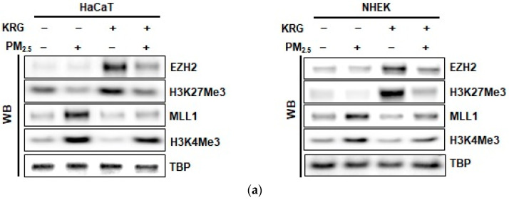
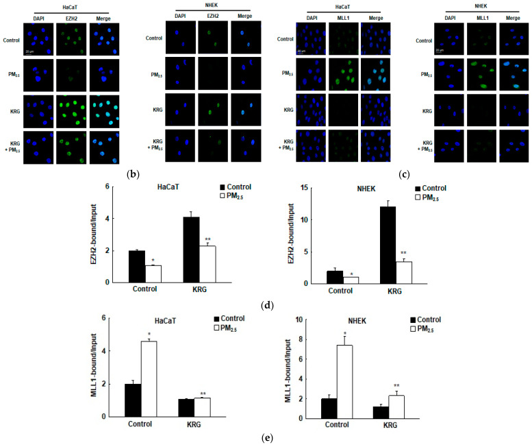
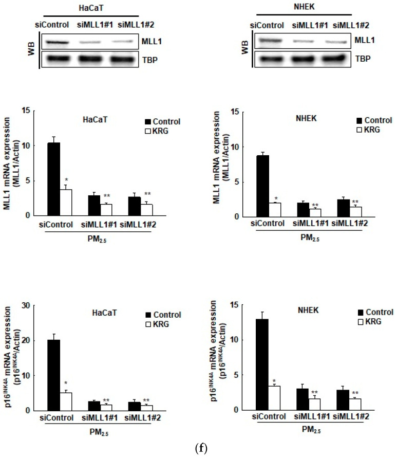
Figure 4.
Attenuation of p16INK4A expression in KRG-treated cells via changes in histone methylation. (a) Nuclear fractions were electrophoresed, and enhancers of zeste homolog 2 (EZH2), H3K27Me3, mixed-lineage leukemia 1 (MLL1), and H3K4Me3 were detected by western blotting with the corresponding antibodies. As indicated, TBP serves as the loading control. (b,c) The nuclear locations of (b) EZH2 and (c) MLL1 were determined by confocal microscopy after Alexa488-labeling (green) with the corresponding antibodies and staining with DAPI (blue). (d,e) ChIP assays using antibodies against (d) EZH2 and (e) MLL1 were performed and analyzed by qPCR. MLL1 siRNA was transfected into cells and incubated for 24 h. (f) Nuclear fractions were electrophoresed, and MLL1 was detected by western blotting with the corresponding antibodies. As indicated, TBP serves as the loading control. mRNA levels of MLL1 and p16INK4A were detected by qRT-PCR. * p < 0.05 and ** p < 0.05 indicate significant differences with control cells and PM2.5-treated cells, respectively.


