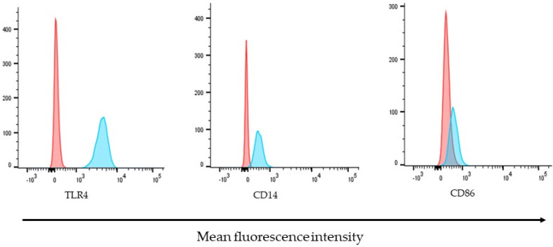
Figure 3.
Molecules of immunological importance on the surface of ARPE-19 cells. The expression of TLR4, CD14, and CD86 were determined by their detection, with specific antibodies coupled to different fluorochromes. Histograms are representative of three independent assays and show the relative fluorescence intensity of ARPE-19 cells, unstained control (red) or stained (blue) with APC-conjugated-Anti-TLR4, FITC-conjugated-anti-CD40, or PE-conjugated-CD86 antibodies.

