Figure 1.
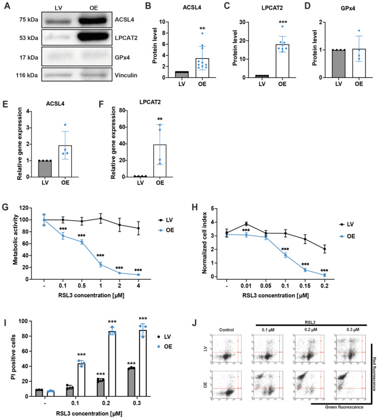
Quantification of protein and mRNA levels and sensitivity of empty vector control and ACSL4/LPCAT2 overexpressing HEK293T cells against RSL3. (A–D) Overexpression of ACSL4 and LPCAT2 by the sleeping beauty system was confirmed by (A) Western blot analysis, which was quantified for protein levels (B) ACSL4 (n = 10) (C) LPCAT2 (n = 8) and (D) GPx4 (n = 4) and depicted as fold protein level normalized to controls. (E,F) Quantitative PCR analysis of untreated LV and ACSL4/LPCAT2 OE cells. Relative gene expression was calculated for (E) ACSL4 and (F) LPCAT2 by normalization to GAPDH RNA level and LV control conditions (data are given as individual data points ± SD; n = 4 replicates per group). Sensitivity against RSL3 was analyzed by (G) MTT assay after 16 h of RSL3 treatment (percentage of control condition) and (H) xCELLigence real-time impedance measurement evaluated 19 h after treatment onset. Data are given as mean ± SD (n = 8 replicates). (I) Cell death was quantified by FACS analysis of PI staining after 16 h of RSL3 treatment (5000 cells per replicate of n = 3 replicates, percentage of gated cells). (J) Representative dot plots of the FACS measurements. *** p < 0.001; ** p < 0.01 compared to (untreated) control (ANOVA, Scheffé’s test).

