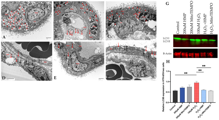
Figure 7.
Autophagy and mitochondrial dysfunction in human pregnancy and in HTR-8/Svneo cells. Electron microcopy images of human placental villous tissue from preeclamptic (A–C) and normal pregnancies (D–F). Red arrows depict lysosomal autophagy structures which are significantly increased in the preeclamptic syncytiotrophoblast cells compared to control villous tissue. Mitochondria (black arrows) are better preserved in syncytiotrophoblast cells from control pregnancies. (Scale bar: 2 µm) (SCT: syncytiotrophoblast, CT: cytotrophoblast, End: endothelium and Nucl: nucleus). (G) Western blot analysis of autophagy markers: LC3B-I and -II (LC3-I and LC3-II in HTR-8/SVneo cells incubated with 100 μM H2O2 for 20 h in the presence of 200 μM HMP or 200 μM MitoTEMPO. The expression of beta-actin was used as an internal control. (H) Densitometry analysis of Western blot shown in (G). These experiments were independently performed at least three times (composite result shown in Supplementary Figure S4). Mann–Whitney-U test, median [IQR]. Control vs. 100 μM H2O2: **: p < 0.01, 100 μM H2O2 vs. H2O2 + HMP: **: p < 0.01 and 100 μM H2O2 vs. H2O2 + MitoTEMPO: **: p < 0.01.

