Figure 5.
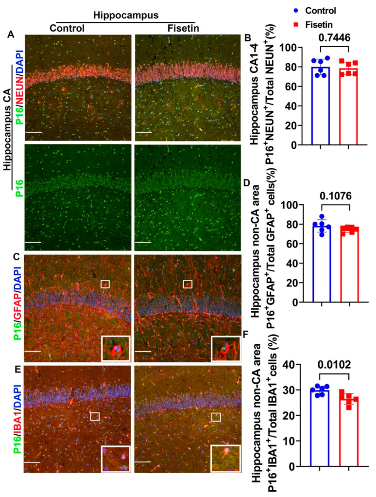
Effects of fisetin treatment on the cellular senescence of different cells in the hippocampus. (A) NEUN and P16 double immunofluorescence staining for the hippocampus, focusing on the CA area. NEUN+ cells are stained in red and many are colocalized with P16 (merged channel). P16+ cells are stained in green in the nuclei (green channel). (B) Quantification of P16+/NEUN+/Total NEUN+ cells in the CA1-4 area. (C) GFAP and P16 double immunofluorescence staining in the hippocampus. GFAP+ cells are stained in red, showing many projections, and are mainly located in the non-CA area. P16+ cells are stained in green in the nuclei. (D) Quantification of P16+GFAP+/Total GFAP+ cells in the non-CA area of the hippocampus. (E) IBA1 and P16 double immunofluorescence staining in the hippocampus. IBA1+ cells are stained in red and mainly located in the non-CA area. P16+ cells are stained in green in the nuclei. (F) Quantification of P16+/IBA1+/Total IBA1+ cells in the non-CA area of the hippocampus. White boxes in each image highlight positive cells of each staining. Scale bars = 100 µm. Exact p values are indicated between the group bars.

