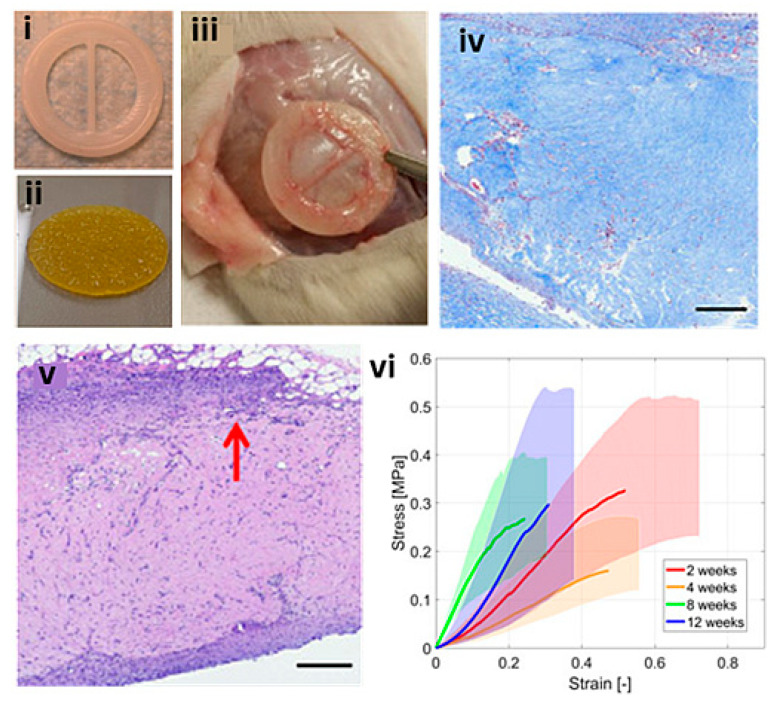
Figure 12.
Simplified bioplotted valve made from (i) PCL frame and (ii) bioplotted type I collagen hydrogels and rMSCs, with (iii) showing subcutaneous explanation from Sprague–Dawley rat after 12 weeks, (iv) H&E staining showing increase in host cellular concentration found at the periphery (red arrow) and infiltrating within the scaffold, (v) Masson’s trichrome showing a diffuse blue expression representative of collagen, scale bars = 300 μm, (vi) stress–strain plot of the heart valve scaffolds at 2, 4, 8, and 12 weeks. Reprinted with permission under the open access from Maxson et al. [150].

