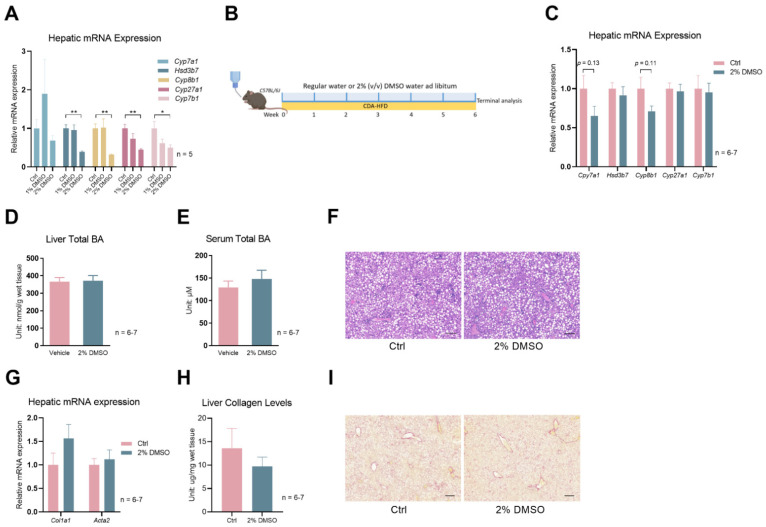
Figure 4.
DMSO does not protect mice from CDA–HFD-induced liver fibrosis. (A) Expression of Cyp7a1, Hsd3b7, Cyp8b1, Cyp27a1, and Cyp7b1 in WT mice kept on 1% or 2% (v/v) DMSO water for one week; n = 5 for each group. (B) Schematic of mouse treatment for panels (C–I): C57Bl/6J WT mice were all treated with CDA–HFD, but with or without 2% (v/v) DMSO in their drinking water. The treatment lasted for 6 weeks. (C) Hepatic expression of Cyp7a1, Hsd3b7, Cyp8b1, Cyp27a1, and Cyp7b1 in mice described in Panel (B). (D) Total BA levels in the liver. (E) Total BA levels of serum. (F) Representative H&E staining of liver sections from mice described in panel (B). Scale bar: 200 µm. (G) Hepatic expression of Col1a1 and Acta2. (H) Liver collagen levels in the mice described in panel (B). (I) Representative Sirius Red staining of liver sections from mice described in panel (B). Scale bar: 200 µm. For panels (C–I), n = 7 for the control group and n = 6 for the 2% DMSO group. All data are mean ± SEM; * p < 0.05, ** p < 0.01, relative to the control group.

