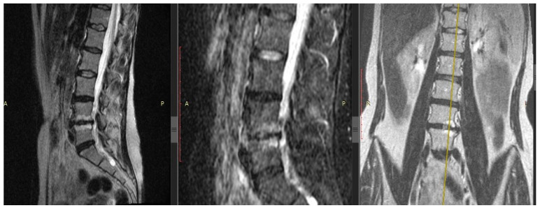
Figure 1.
Patient with back pain. Magnetic resonance imaging of the lumbar spine, modes T2-weighted images with fat suppression in sagittal sections (left and center images, respectively), T2-weighted short-tau inversion-recovery image in the coronary section (right image). Right-sided lumbar scoliosis, the 5th stage of degenerative disc disease by Pfirrmann at the L4/5 and 4th stage—at the L5/S1 level, with IVD hernias L4/5 and L5/S1, erosion of the endplates and Modic changes type 1 in the vertebral bodies L4/5 (yellow axis carried out via Modic-1). Modic-1 = the bone marrow edema; Modic-2 = the bone marrow fatty degeneration.

