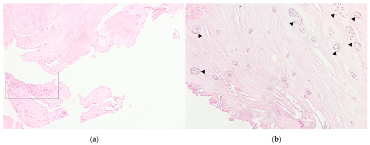
Figure 2.
Patient intervertebral disc sample obtained as a result of microdiscectomy in the patient in her 40 s, with a hernia of the L5/S1 vertebrae. Own data, 2020. Hematoxylin–eosin staining. (a). Light microscopy, magnification 100×. Zones of inflammatory infiltration, combined with vascularization and granulation tissue. (b). The sample of the same patient, magnification 100×. Clusters of nucleus pulposus cells (triangles) are a characteristic sign of disc degeneration.

