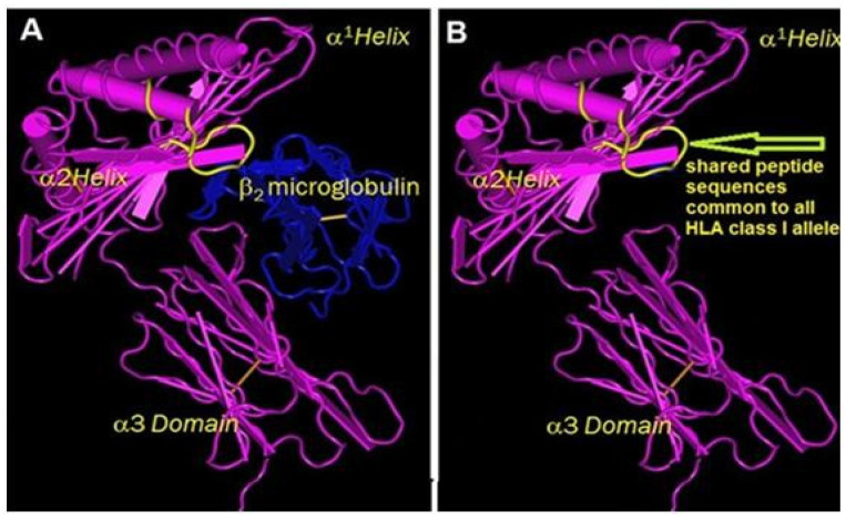
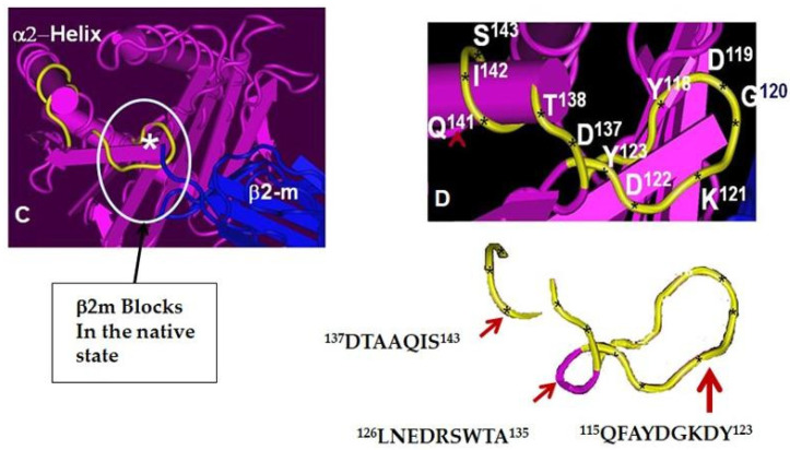
Figure 3.
(A,B) Structure of HLA-I intact molecule (Face-1) showing sequences shared by all HLA loci (yellow string in α2 helical domain), which is masked by B2M (shown in blue). (C,D). The position of most commonly shared sequences are as follows: 137DTAAQI142 (present in HLA-B and HLA-C in G-ALPHA2 between the D strand and the helix, IMGT numbering 49–54, Lefranc et al. [3]) and 115QFAYDGKDY123 (present in all loci of HLA-Ia and Ib in G-ALPHA2 BC loop, IMGT numbering 25–33). The binding of mAb MEM-E/02 to HLA heavy chain coated solid matrix is inhibited by the above-mentioned peptides, Ravindranath et al. [37], shown as yellow curved line. In Face-1, these peptides are masked by B2m, but exposed in B2m-free HCs. The asterisk in figure show the sequence blocked by B2m in the native state.


