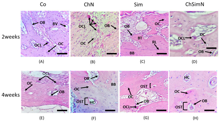
Figure 4.
Histological section of the bone defect. (A) Control group (Co) at two weeks, showing osteoblasts (OBs), blood vessels (BVs), osteoclasts (OCLs) and osteocytes (OCs). (B) Chitosan nanoparticle group (ChN) at two weeks, showing multiple OCLs, OBs and OCs. (C) Simvastatin group(Sim) at two weeks, showing basal bone (BB) separated from the new bone trabeculae (BT) by a reversal line (RL), OCs and OBs. (D) Combination of chitosan nanoparticles and simvastatin(ChSimN) at two weeks, showing OCLs, OBs on the border of the new trabeculae and OCs embedded in the new bone. (E) Co at four weeks, showing mature bone trabeculae containing OCs, OBs on the border and OCLs. (F) ChN group at four weeks, showing OCs arranged in a circular pattern around the haversian canal (HC) forming the osteon (OST), OBs and RL separating the BB from the new bone. (G) Sim group at four weeks, showing OST, OBs and OCs. (H) ChSimN group at four weeks, showing OST, HC, OBs and OCs. H&E × 40. Scale bar = 50 µm, n = 42.

