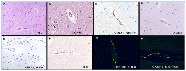
Figure 8.
Histologic and molecular correlates of COVID-19 in human brains. Panel (A) shows the microvessels in normal brain. In comparison, many of the capillaries in COVID-19 brain tissues show marked perivascular oedema (panel (B)). Serial section analyses of the COVID-19 brain shows that the endothelial cells of the microvessels contained the spike glycoprotein (panel (C)), the ACE2 receptor (panel (D)) and IL 6 (panel (F)), but not viral RNA (panel (E)). The fluorescent yellow signal marks co-localisation of the spike protein with IL6 (panel (G)) and caspase 3 (panel (H)), respectively, in these endothelial cells. Each magnification is 800× with DAB (brown) signal (panels (C–F)) or Fast Red (red) (panel D). (For interpretation of the references to colour in this figure legend, the reader is referred to the web version of this article.). Reprinted from Annals of Diagnostic Pathology, Vol. 51, Nuovo GJ, Magro C, Shaffer T. et al., Endothelial cell damage is the central part of COVID-19 and a mouse model induced by injection of the S1 subunit of the spike protein. Figure 1, 151682, Reprinted with permission from Ref. [243]. Copyright (2020) Elsevier.

