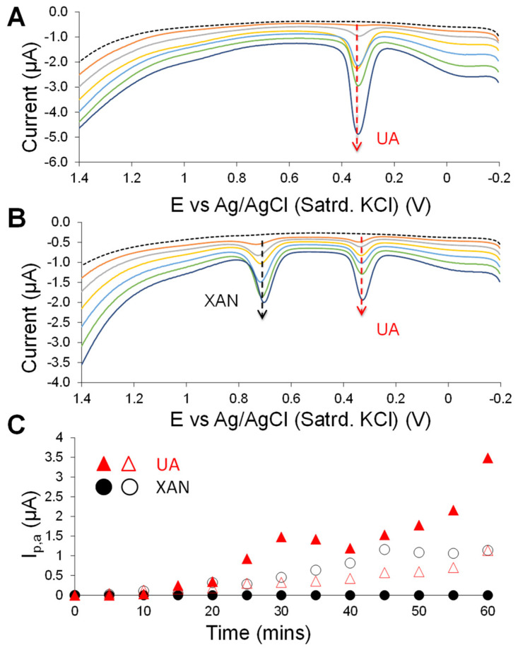
Figure 4.
Sequential DPV scans over time before (dashed trace) and after (solid traces) exposure to 200 µM XAN (10 mM PBS; pH 7) at GCEs modified with C6-MPC-doped HMTES xerogels (with XOx) either (A) with or (B) without the PU capping layer (scans every 10 min shown); (C) oxidative, anodic peak currents (Ip,a) measured from the previous DPV results (A,B) at +0.380 V for XOx generated UA and +0.725 V from injected XAN at C6-MPC-doped HMTES systems with (solid markers) and without (open markers) PU capping layers measured over the course of 1 h.

