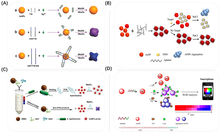
Figure 7.
(A) Schematic illustration of the presented sensing strategy for the colorimetric detection of Hg2+ based on the growth of Au NSPs induced by different amounts of bases. The black line represents the binding domain, and the blue line represents excess bases except for the binding domain. Reproduced with permission from [84]. Copyright 2017, Wiley. (B) Principle of quantitative colorimetric detection of SDM based on Au NSPs aggregation with a smartphone. Reproduced with permission from [86]. Copyright 2022, Elsevier. (C) Schematic illustration of the proposed SMBs–Apt1 sandwich-based colorimetric sensor for detection of S. typhimurium in milk (Size not to scale). Reproduced with permission from [87]. Copyright 2020, Elsevier. (D) Schematic illustration of the detection of TET/CAP based on Au NSP colorimetric aptasensors. The Apt acts as a molecular switch adjusting the Au NSP aggregation. When antibiotics remove the fragment of Apt from the Au NSPs’ surface, unbalanced Au NSPs are aggregated on different scales under high-salt conditions. It thereby causes colloidal color changes, which can be detected by UV spectroscopy and Smartphone analysis, respectively. Reproduced with permission from [70]. Copyright 2020, Elsevier.

