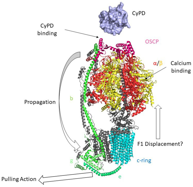
Figure 3.
Mammalian monomeric F-ATP Synthase and the “Death Finger” Hypothesis of mPTP formation. Cryo-EM structure of F-ATP Synthase is shown (PDB ID: 6TT7 [101]). According to the death finger hypothesis, calcium binding to the α (red)/β (yellow) subunits induces conformational changes that are transmitted from OSCP (pink), the binding site of CyPD (light blue, PDB ID: 2Z6W [54]) to the lateral stalk of F-ATP synthase (subunit b in light green) up to the g (lime) and e subunits (dark green). This would lead to the removal of lipids within the c-ring (cyan) and the displacement of the F1 sector from the Fo, leading to mPTP opening.

