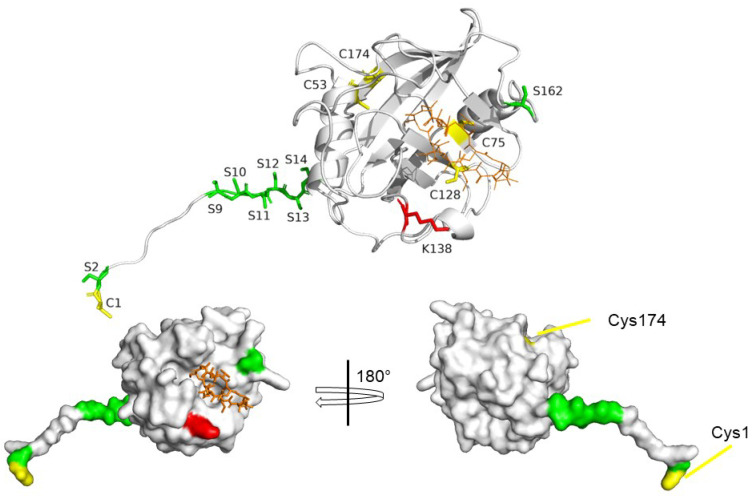
Figure 4.
Post-Translational Modification of CyPD. Cartoon (upper panel) and surface (lower panel) representation of CyPD (AlphaFold structure) active site side (upper panel, lower left panel) and back face side (lower right panel) and PTM target sites. Red: acetylation site. Yellow: cysteine residues. Green: serine residues involved in protein phosphorylation.

