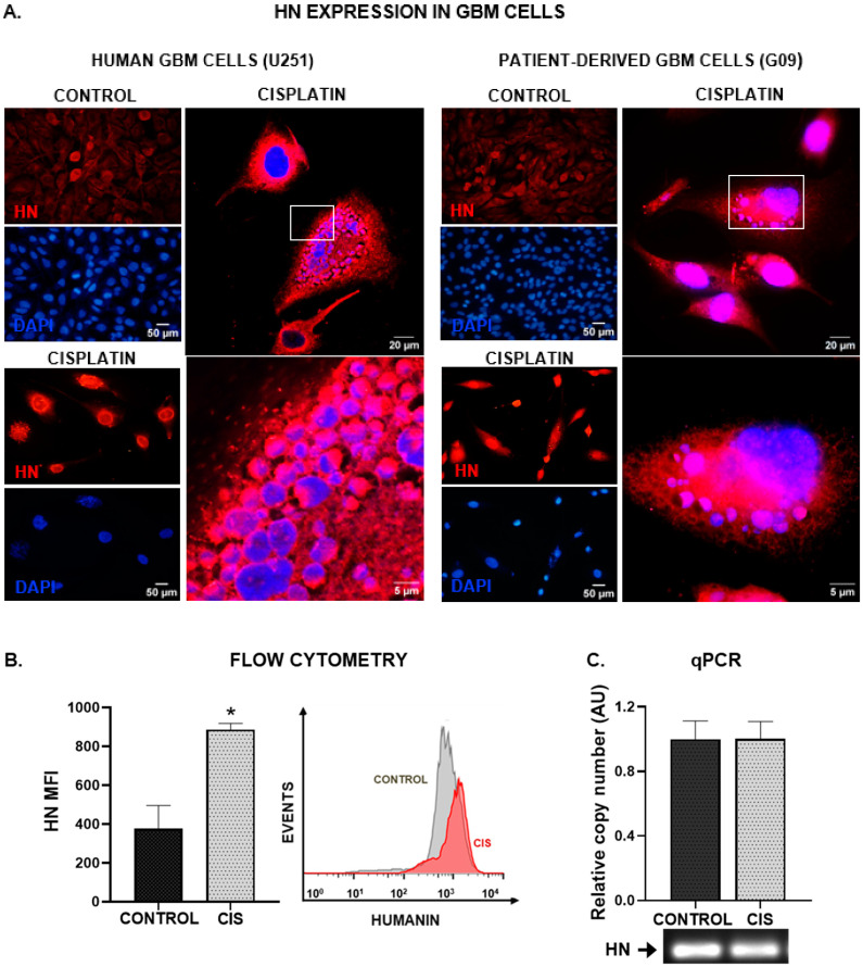
Figure 1.
Chemotherapy upregulates HN in human GBM cells. Human U251-MG GBM cells and patient-derived GBM cell cultures (G09) were incubated with 5 µM cisplatin for 48 h. HN expression was assessed by immunofluorescence (A), flow cytometry (B) and qPCR (C). (A) Images show cells immunostained with HN antibody (red), and DAPI-stained nuclei (blue). Representative magnified images of cisplatin-treated human GBM cells were obtained by confocal microscopy. The white box indicates the area magnified in the bottom panel. (B) Mean fluorescence intensity (MFI) of HN staining in human U251-MG GBM cells (n = three replicates/condition). A representative histogram is depicted. * p < 0.05, Student’s t test. (C) Expression of HN mRNA as assessed by qPCR. A representative gel of qPCR products is shown.

