Figure 3.
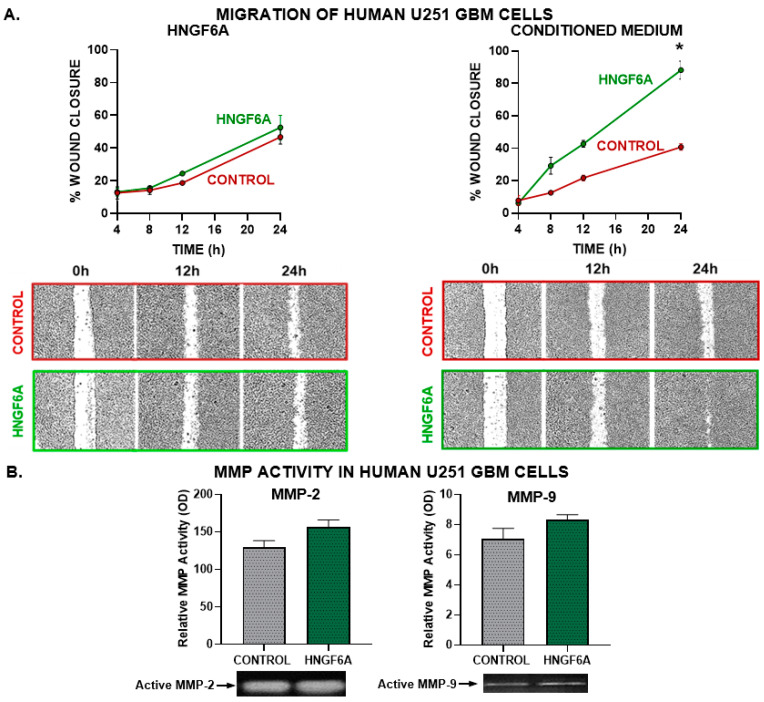
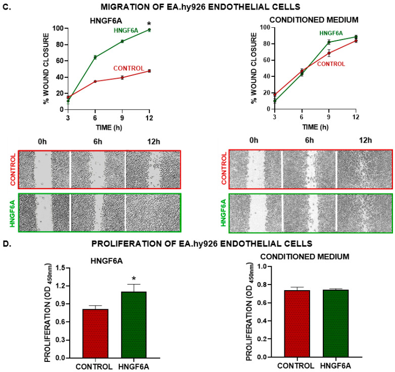
Effect of HNGF6A on GBM cell migration and angiogenic capacity. (A) GBM U251-MG cells were seeded to confluence and incubated with HNGF6A (1.25 μM, left panels) or with conditioned media from HNGF6A-treated cells (right panels). The monolayer was scratched and the cell-free area was measured at different time points. * p < 0.05 (nonlinear regression analysis). Representative images of the scratch areas are shown. (B) SDS-PAGE gelatin zymography of conditioned media from human GBM U251-MG cells incubated in the presence of HNGF6A (1.25 μM) for 48 h. The bands were analyzed by densitometry with the ImageJ software (Version: 1.53k) and the zymographic activity was expressed as a percentage in relation to a standard internal sample that is saturated at a density of 50%. * p < 0.05 Student’s t-test (C) EA.hy926 endothelial cells were seeded to confluence and incubated directly with HNGF6A (1.25 µM) or using conditioned media from HNGF6A-treated U251-MG cells. A scratch test was performed and the cell-free area was measured at different time points. * p < 0.05 (nonlinear regression analysis). (D) EA.hy926 endothelial cells were incubated with HNGF6A (1.25 µM) or with conditioned media from HNGF6A-treated U251-MG cells for 48 h and proliferation was determined by BrdU incorporation (ELISA) * p < 0.05 Student’s t-test.


